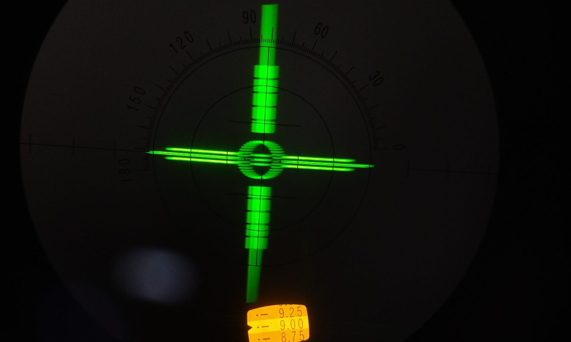
This is a question-answer page, to help people better understand glaucoma

Contents:
- What is glaucoma?
- What do you see in glaucoma – or what can no longer be seen?
- Why did glaucoma develop in my eyes?
- Does vision worsen rapidly?
- What is a normal eye pressure?
- How is glaucoma treated?
What is glaucoma?
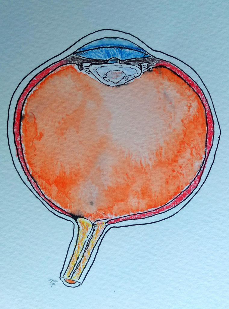
This is an eye cut in half:
Glaucoma is a collective name given to several eye conditions which progressively or gradually damage the optic nerve. This is the cable-like structure at the back of the eye (circled in blue below).
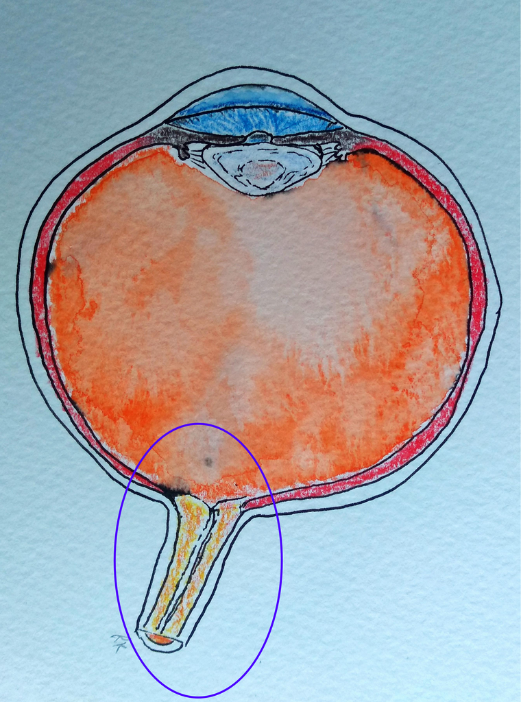
The optic nerve carries visual information from the eye to the brain for further processing. It is itself part of the brain, so like other forms of brain damage, damage to the optic nerve is usually permanent and does not recover.
In some cases, the eye pressure is higher than average, but in other cases the pressure seems within to be at a normal level. Treatment to reduce the eye pressure can halt worsening of glaucoma. This is why eye pressure is an important thing to measure in this disease.
What do you see in glaucoma – or what can no longer be seen?
As a result of glaucoma, areas within the field of vision begin to disappear gradually. This change usually occurs without symptoms such as pain or blurred vision. The brain compensates for the missing areas by using the vision from the other eye and by filling in the view with its own creations, which is why the changes can be difficult to notice yourself.
This can sometimes lead to difficulties with reading or moving around. Reading may become challenging if the visual field is not wide enough to allow the eyes to scan across text from left to right. If visual field defects are present in both eyes, a driving licence may be at risk. Losing a driving licence can significantly affect quality of life and everyday functioning.
In general, glaucoma does not cause black shadows in the visual field, and people with glaucoma usually do not report that they are “seeing through a tunnel.” The brain is surprisingly good at filling in missing parts of the visual field. Very often, even when the visual field is already so damaged that driving is no longer legally permitted, a person with glaucoma may still be convinced that their vision is completely normal. Most often, if the field of vision has been damaged, people report noticing missing areas or blurred areas (this has been expertly studied by David Crabb and his research group https://doi.org/10.1016/j.ophtha.2012.11.043).
Why did glaucoma develop in my eyes?
In most cases in Finland and Europe, the exact cause is not known. However, glaucoma appears to be related to impaired drainage of the fluid in the eye’s anterior chamber, the internal front part of the eyeball. This front part of the eye is shaped a little like the building formerly known as the Millenium Dome. The roof of this structure is like the eye’s cornea, the see-through window right at the front of the eye.
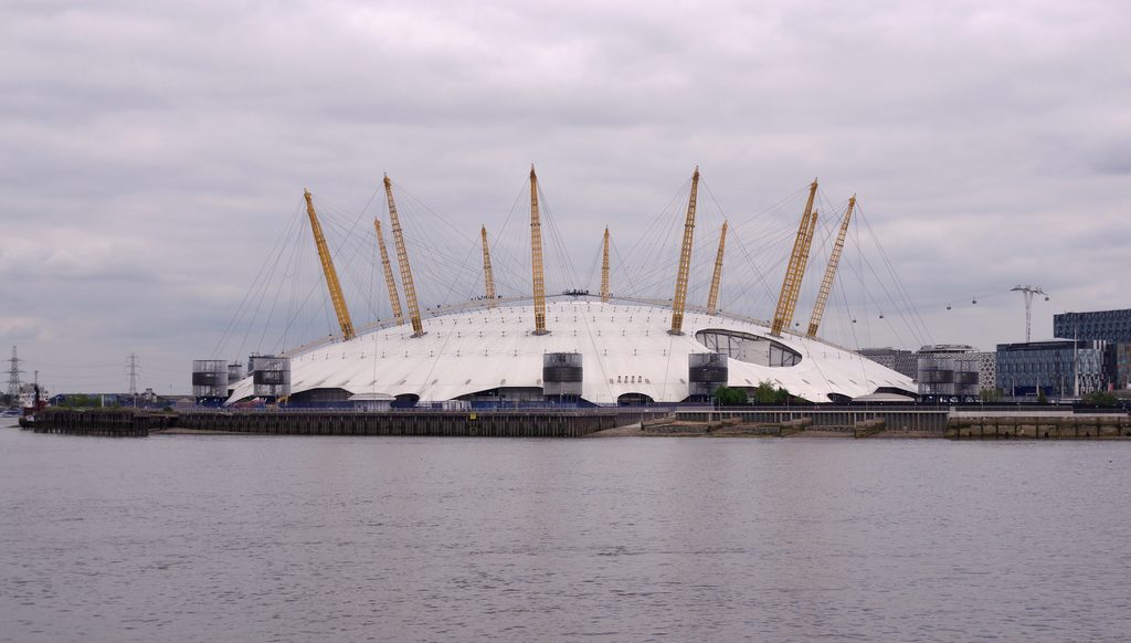
Fluid exits the eye through exit channels at the base of this dome, which is known as the “drainage angle”, just as people leave this arena through doors at the base of the structure. The part of the angle through which fluid goes is called the trabecular meshwork.
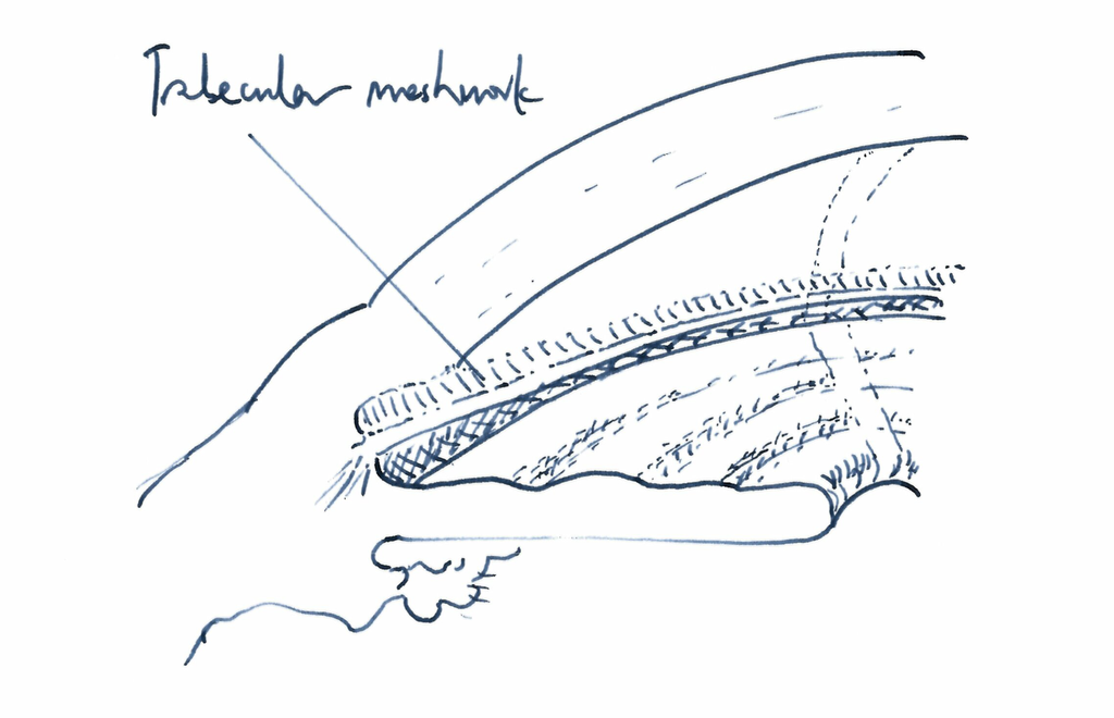
From there, the fluid from the eye goes into veins and back to the heart.
This is not related to the drainage of tears, which occurs outside the eye through the eyelids into the tear ducts and then into the nose.
If the drainage of fluid produced inside the eye is reduced, the pressure inside the eye may rise. A significant part of glaucoma risk is hereditary, that is, the tendency to develop the disease can run in families, but this usually cannot be explained by a single gene. It is likely that several different genes are involved, but their interactions are not yet fully understood.
In some glaucoma cases, the disease is caused by differences in the internal structure or shape of the eye.
In other glaucoma cases, the disease is caused by another condition affecting the eye, such as inflammation (iritis), previous eye surgery, medications currently in use, or an eye injury.
Does vision worsen rapidly?
Usually no. If the intraocular pressure is not significantly high, glaucoma often worsens slowly over years, sometimes even though eye-pressure lowering treatment has not been used.
With proper monitoring and treatment, most eyes with glaucoma do not worsen, or undergo very slow visual field deterioration.
However, if the eye pressure is very high or if the glaucoma is caused by another eye condition, the deterioration of the visual field can occur quickly — even within a few weeks or months. In these types of glaucoma, eye pressure should be monitored more frequently than usual. For example, an inexpensive pressure check every 1–2 months at a nearby optician can reduce the risk of progression between visits to the ophthalmologist.
What is a normal eye pressure?
A normal eye pressure is usually around 23 mmHg or below when measured with a reliable device. This value is used in national referral guidelines in several countries.
“Normal” eye pressure is based on average values measured in the general population, and it is not an absolute boundary between healthy and diseased eyes.
In an English study, about 76 percent of people diagnosed with new primary open‑angle glaucoma had an average eye pressure below 21 mmHg.
For this reason, the concept of “normal eye pressure” can be misleading.
A more important question is: what is a healthy eye pressure for my right and left eye specifically? This target pressure may be different in each eye. The goal of treatment is not to reach a certain population‑defined normal value, but to find an eye pressure at which glaucoma does not cause further damage to the optic nerve
How is glaucoma treated?
The most common initial treatment for glaucoma is either laser therapy performed during a clinic visit or starting eye drops. The goal of these treatments is to lower the eye pressure and thereby prevent progressive visual field damage. Eye drops usually need to be used indefinitely, because once they are stopped, the eye pressure rises back to a harmful level.
The laser treatment is called SLT (selective laser trabeculoplasty). Its exact mechanism of action is not fully understood, but the treatment is very safe when performed on eyes in which the structure of the eye’s drainage channel is clearly normal. The treatment can also sometimes be performed on other eyes, as long as necessary precautions are taken.
However, the laser does not always succeed in lowering eye pressure, and the duration of its effect varies: if the treatment works, the effect may last from months to years.
In cases where glaucoma is already advanced at an early stage, or where there is a risk that the visual field will worsen despite laser or medication treatment, surgery may be considered. This is discussed in more detail on another page.